

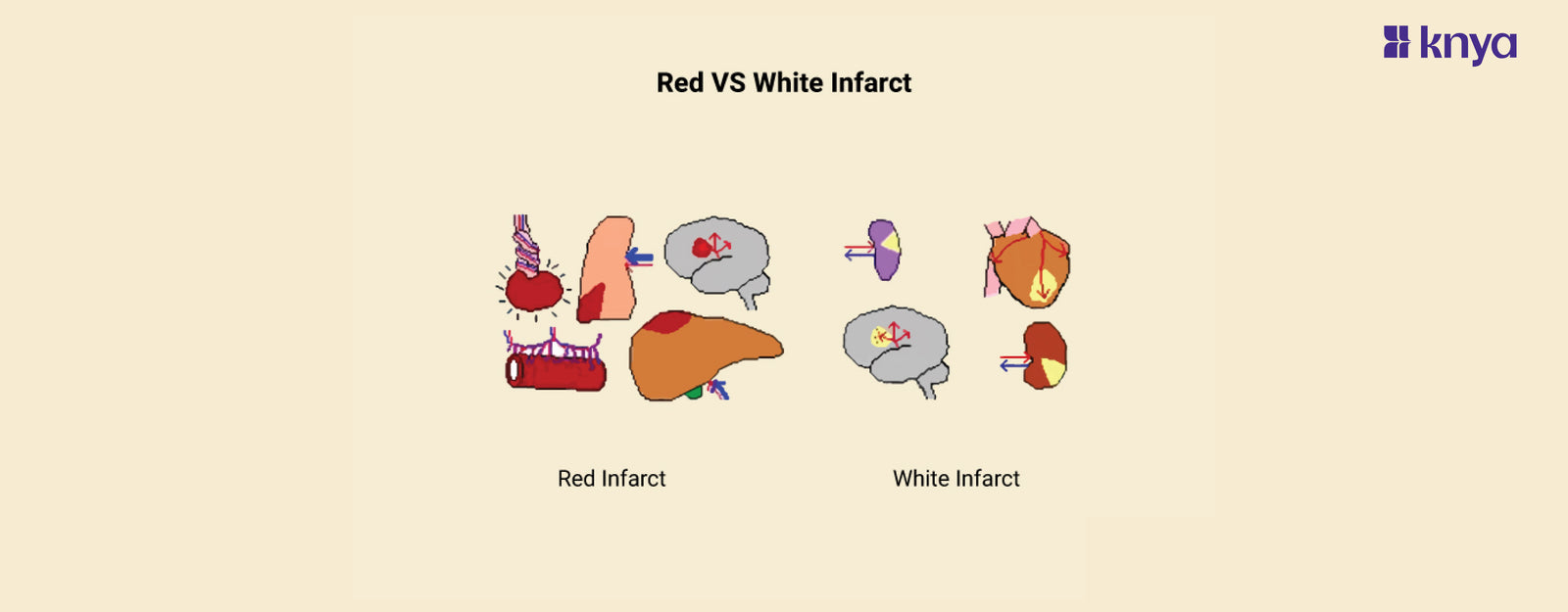
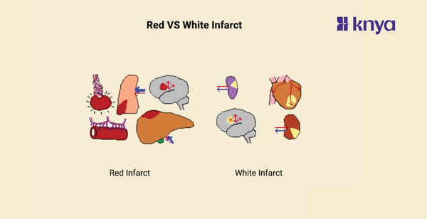


Difference Between Red and White Infarct: Red and white infarcts are both areas of tissue death due to insufficient blood flow, but they differ in appearance and underlying cause. Red infarcts, also known as hemorrhagic infarcts, are usually red or brown due to bleeding into the dead tissue. They occur in organs with loose networks of blood vessels or dual blood supplies, like the lungs or intestines, when a vein is blocked. White infarcts, also called anemic infarcts, appear pale due to the lack of blood. They often affect solid organs with single blood supplies, like the heart or kidneys, when an artery is blocked. Understanding the difference between red and white infarcts helps with diagnosis and treatment, as they involve different causes and may require different interventions. Remember, this is just a simplified explanation, and it's crucial to consult a medical professional for accurate diagnosis and treatment advice.
Difference Between Red and White Infarcts
Red infarct and white infarct are two types of tissue infarctions that occur due to inadequate blood supply to a particular area of tissue, leading to ischemia and necrosis. Here are the key difference between the two:
|
Feature |
Red Infarct |
White Infarct |
|
Color |
Appears red due to hemorrhage and blood extravasation into the necrotic tissue. |
Appears pale or white due to lack of blood supply and subsequent coagulative necrosis without significant hemorrhage. |
|
Cause |
Typically occurs in tissues with dual blood supply or where there is loose connective tissue allowing blood to pool. |
Occurs in solid organs with end-arterial blood supply. |
|
Type of Necrosis |
Liquefactive necrosis due to the presence of blood. |
Coagulative necrosis. |
|
Common Sites |
Lungs, intestines, testes. |
Heart, spleen, kidney. |
|
Consistency |
Soft consistency due to liquefactive necrosis and hemorrhage. |
Firm consistency due to coagulative necrosis. |
|
Associated Conditions |
Venous occlusion or in tissues with a rich collateral blood supply. |
Arterial occlusions. |
|
Microscopic Appearance |
Erythrocytes extravasated into the necrotic tissue. |
Coagulative necrosis with preservation of tissue architecture. |
|
Clinical Implications |
May lead to hemorrhagic complications. |
May result in more localized tissue damage without significant hemorrhage. |
|
Reversibility |
Higher chance of reversible damage if blood flow is restored promptly. |
May lead to irreversible tissue damage more quickly. |
|
Risk Factors |
Conditions like thrombosis or embolism with reperfusion. |
Atherosclerosis and thromboembolism causing arterial occlusions. |
Browse Best Scrubs Collection
What are Red infarcts?
Red infarcts, also known as hemorrhagic infarcts, occur when a vein (blood vessel carrying blood away from the heart) is blocked, causing blood to leak into the dead tissue, giving it a reddish appearance. These typically affect loose organs like lungs, intestines, and testes, with dual blood supplies or congested tissues. They often arise from vein blockage, severe vasoconstriction, or reperfusion injury after a previously blocked artery is reopened.
Key Features of Red infarcts:
- Cause: Typically caused by venous occlusion, where blood flow is blocked on the way out of the tissue.
- Appearance: Reddish due to blood accumulation in the dead tissue.
- Location: Commonly seen in loosely packed organs like lungs, intestines, and testes.
- Example: Hemorrhagic stroke, which occurs when a blood vessel in the brain ruptures and bleeds into the surrounding tissue.
What are White Infarcts?
An artery (blood vessel supplying blood to the heart) obstruction causes white infarcts, also known as anaemic infarcts, which deprive tissues of oxygen and nourishment. Solid organs such as the kidneys, spleen, and heart suffer from cell death and appear pale as a result, frequently with no other blood supply. Inflammation, cholesterol accumulation, and blood clots are common causes of arterial occlusions.
Key Features of White Infarcts:
- Cause: Typically caused by arterial occlusion, where blood flow is blocked on the way into the tissue.
- Appearance: Whitish or pale due to lack of blood.
- Location: Commonly seen in solid organs like heart, kidneys, and spleen.
- Example: Myocardial infarction (heart attack), which occurs when a coronary artery is blocked, depriving the heart muscle of oxygen and nutrients.
Shop Best Lab Coats from Here!
Similarities Between Red and White Infarcts
- Red and white infarcts result from ischemia, which is inadequate blood supply to a tissue.
- Both types of infarcts involve tissue necrosis, leading to loss of function in the affected area.
- Both types of infarcts may present with similar clinical symptoms depending on the organ affected, such as pain, organ dysfunction, or systemic symptoms like fever or leukocytosis.
- Both red and white infarcts can be identified through histological examination of affected tissues, revealing characteristic changes associated with ischemic necrosis.
- Both types of infarcts may lead to complications such as inflammation, infection, or organ dysfunction if not promptly managed.
Even though they both show tissue death from blood flow restriction, red and white infarcts depict somewhat distinct images. Leaking blood cells give red infarcts their crimson appearance. These are frequently observed in organs with looser architecture and multiple blood supply, such as the lungs and intestines. Usually, reperfusion damage or venous obstructions cause them. White infarcts, on the other hand, are less red in colour due to restricted blood seepage and are frequently observed in solid organs such as the kidneys and heart that have a single blood supply. These result from arterial occlusions depriving the tissue of nutrition and oxygen. Recognising these colour differences emphasises the critical function that blood flow dynamics play in tissue health and helps with diagnosis and therapy.
| Check out More Articles | |
| Difference Between Tendon and Ligament | |
| Difference Between Seizure and Epilepsy | |
| Difference Between Hypothyroidism and Hyperthyroidism | |